
New research from Mount Sinai suggests newly developed tests could help assess traumatic brain injury (TBI) in injured patients on and off the battlefield.
As we discussed earlier, TBI occurs when the brain is subjected to a hit or event that causes torsion, shearing, or bruising within delicate brain tissue. Nerve cells are damaged and may collapse or leak chemicals into the brain, compounding the injury.
The ability to diagnose TBI is crucial to short-term treatment and the long-term prognosis of a patient. Brain injury can cause changes that trigger anxiety, depression, personality shifts and neurodegenerative conditions that include dementias, Alzheimer’s disease, and Chronic traumatic encephalopathy (CTE).
A blood test has long been sought to assist with diagnosis and replace radiation-intensive diagnostic CT scans. In a study published recently in Molecular Psychiatry, researchers took a two-fold approach to advance diagnostic capabilities after a TBI.
From the battlefield and the research lab
Looking to veterans returning from combat in the Middle East, research scientists used imaging, neurocognitive and neuropsychological assessments, and blood biomarkers to evaluate chemical and structural changes that occur in the brain after injury.
The veterans involved in the study each reported between one and 50 blast exposures and all complained of chronic cognitive and behavioral changes. Alongside evaluation of human patients, researchers used mouse models to compare traits, functional, and chemical consequences that occur after TBI.
Some key points of the study include the following:
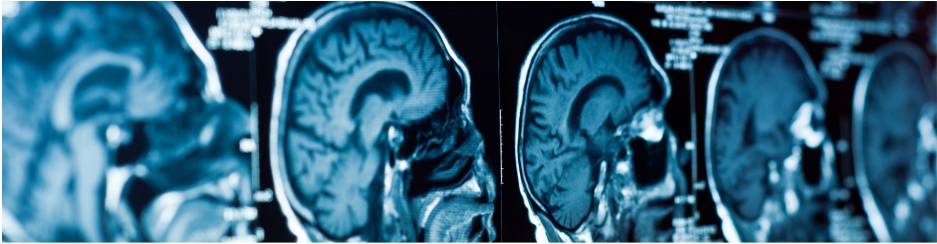
- In a healthy brain, a protein called tau helps form the internal framework of nerve cells in the brain. After injury, or repetitive injury, tau may begin to clump together, causing build-up that collapses the cell and forms twisty tau chains that create additional brain damage. In this study, researchers were able to use positron emission topography (PET) scan to locate excessive tau bundles in five of the ten veterans who took part in the study.
- Neurofilament protein-light chain (Nf-L) is a brain protein that leaks from brain tissue after a traumatic brain injury. Because of this leakage, researchers were able to measure its presence in the blood. Focus on Nf-L allowed the scientists to identify altered blood levels of the protein after mild TBI, in veterans suffering chronic effects of brain injury, and in others with neurodegenerative diseases.
Research team member Dr. Gregory Elder of Mount Sinai remarks, “There are many young, otherwise healthy veterans who have suffered blast-related TBIs, some of them years in the past, who either aren’t getting better or, in some cases, are getting worse. We don’t know why or how to identify those at greatest risk. The work in this study is a step towards answering those questions.”
This study contributes refined diagnostic knowledge along with better understanding of the neurodegenerative changes that occur in the human brain after an injury, wherever it occurs.
Skilled medical malpractice attorneys help you in Maryland and Washington, DC
With more than 35 years of successful experience obtaining more than one billion dollars for our clients, Schochor, Staton, Goldberg, and Cardea, P.A. is dedicated to helping those personally injured through medical malpractice. Call 410-234-1000 or contact us to set up a free consultation to discuss your case.